What is self-cure acrylic resin? best 1

Self-cure acrylic resins have been used mainly in prosthetic dentistry for the construction of custom-built impression trays, occlusion registration bases and for repairing fractured dentures. Recently, pour-type resins have been introduced for the fabrication of denture bases.

self-cure acrylic resin

What are the differences between heat cured acrylic resin and self-cure acrylic resin?

The principal difference between self-cure and heat-cure resins is that more residual monomer is present in the self-cure resins. In addition, with a high monomer to polymer ratio, residual monomer content in the polymerized acrylic resin would be large.

What is heat cured acrylic resin?

Introduction: Heat cure acrylic resins are the most commonly used denture base materials. The important limitation is they may act as reservoir of microorganisms. The adherence of microorganisms can be reduced by chemical modification of the surface charge of denture base resin.

What is cold cure acrylic resin?

Bonding of cold-curing acrylic resin to acrylic resin teeth increases tooth retention and strengthens denture bases because the plastic teeth become an integral part of the denture base. The bonding was verified in experimental dentures made of a compression molded and a pour type poly(methyl methacrylate).

What is acrylic resin used for?

Acrylic resins feature excellent transparency and durability, and are used in a broad range of applications from consumer items like lenses to industrial products like molding materials, coatings and adhesives.

What is denture base resin?

Definition • DENTURE BASE: The part of a denture that rests on the foundation tissues and to which teeth are attached. • RESIN: A broad term used to describe natural or synthetic substances that form plastic materials after polymerization. –

What is a separating medium?

Separating media are those substances which is used to separate two surfaces from each other. Separating media help to separate. A. Plaster surface to acrylic surface.

How do you treat acrylic resin?

In the long curing cycle the acrylic dough was cured at 72 °C for 6.5 h and then 92 °C for 1.5 h. The flasks were left undisturbed in the curing tank for at least 36 h to allow the acrylic resin to cool slowly to the ambient temperature (24–28 °C).

What are acrylic resins used for?

About Acrylic Resins Due to their excellent durability and weatherability as coating materials, acrylic resins are used extensively in applications such as automotive, architectural and plastic coatings.

How long does cold take to cure acrylic?

Figures. curing technique- 15 minutes); G3) cold cure acrylic resin (Ivomat curing technique- 20 minutes). acrylic polymerized chemically on air and under hydraulic press.

Can I use a hair dryer on resin?

Option 3: Turning a hair dryer into an epoxy dryer If there’s one thing resin bubbles can’t stand, it’s the heat. You can actually use a hair dryer to pop bubbles; however, the heat a hairdryer provides is less potent than that of a butane or propane torch.

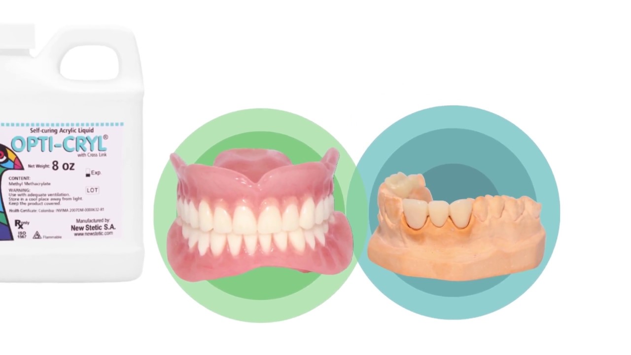
How big is an acrylic monomer heat cure?

Opticryl- Monomer Self Cure only 8oz (Acrylic Resin Liquid) New Stetic veracryl – This Product can only be Shipped by Ground Transportation- it Cannot… Opticryl Dental – Heat Cure Monomer Only 32oz/Quarter Gallon (Acrylic Resin Liquid)…

Which is the best acrylic resin for dental use?

Opticryl Dental – Heat Cure Monomer Only 8oz (Acrylic Resin Liquid) veracryl – This product can only be shipped by ground transportation- it… Dental Combined Organizer, Acrylic Holder Dispenser for Resins, Applicators, Clinic…

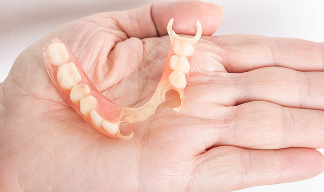
What kind of resin is used for dentures?

Denture acrylic resins are used for the base of dentures. They may be traditional heat-cured, cold-cured or self-cured.

The Pros and Cons of Thermoplastic Partial Dentures

The Pros and Cons of Thermoplastic Partial Dentures

Thermoplastic materials have been available in prosthetic dentistry for more than 30 years and the market place continues to grow with new and existing companies manufacturing their own products.

What makes these dentures different from traditional partial dentures is the material from which they are made and how they are made.

Unlike acrylic dentures, they are made from a thermoplastic nylon resin that is ultra thin, very flexible (think more comfortable for chewing and speaking) and is so durable that one company – Valplast – offers a lifetime warranty for fractures or breaks.

The material doesn’t absorb odors or stains, and if patients suffer from allergies to acrylic or certain metals, it’s a great choice. It contains no BPAs and is considered the most biocompatible material.

Some patients feel that the appliance “disappears” or is “invisible” in their mouth, thus the esthetics of it is far superior to conventional acrylic/metal partial dentures.

The cost may be slightly higher than conventional acrylic partial denture because the fitting and finishing time at the lab is increased, but the result makes it well worth it. 

Thermoplastics like Flexite, TCS (IFLEX) and Valplast all differ from regular thermosets like acrylic powder and liquid because they are already polymerized (cured) when manufactured and shipped to your lab. They can be manufactured in many forms, from sheet to pellets to powder. Once heat is introduced, the plastic is softened to the desired state and then injected into a mold. The only thing that changes is its physical shape; there are no actual chemical changes. Thermoplastics will differ depending on their molecular composition – some require higher temps to become moldable and some require greater injection pressures.

One advantage of this material is that there is also flexibility in the design of the types of clasps. We can often use the circumferential ring clasp on any freestanding tooth and it works well on medially tipped mandibular molars. It can make an excellent transitional restoration during the healing period on implant cases.

Your dental lab will have its preference for which thermoplastic material it prefers to work with. Some are more difficult to finish, fit and adjust than others. Some may require more repairs to the teeth than others. Some may prefer a specific material because of the color blending. A conversation with your technician will help you decide the best material for your patient. Click this link for a video lesson on communicating with your technicians.

The biggest challenge for the dentist is the adjusting and polishing of the material. Do not think acrylic when polishing by using a quick and pressure-applied motion. Think of dividing the appliance in sections and spending one minute per section. That will seem like a long time when your experience is only conventional acrylic resin.

Whether or not you are using Valplast, Flexite, TCS or Iflex, each company has a system for polishing. If you are going to provide these partial dentures, be sure you have the armamentarium to polish them well. 

The partial needs a smooth and satin finish before polishing so use the appropriate rubber wheels.  I typically round the edges of the wheel when I receive them so that I don’t accidentally cut into the nylon with a sharp edge. If there are any spots that the wheel cannot access, I recommend using a Robinson wheel in a horizontal motion with a light dusting technique. 

Patients are very happy with these prostheses because they are fabricated readily and don’t require multiple try-in appointments. I suspect in the future we will make very few traditional metal acrylic removable partial dentures as this material excels for all our patients. The challenge for us is in adjusting it.

Metal Free Crowns

CROWNS SO NATURAL LOOKING YOU’LL THINK THEY ARE REAL TEETH.

A chipped tooth can ruin your appearance and also increase the risk of long-term dental damage. Crowns are the best way to protect your teeth and ensure they’re safe from harm. Metal-free crowns are teeth restorations that are entirely made of ceramic, providing high aesthetic value. The most favourable part about metal-free crowns is that they look very similar to natural teeth, making them the more favoured option today.

WHY ARE METAL-FREE CROWNS RECOMMENDED?

The metal-free crowns available in the dental world today are highly praised for their biocompatible nature. For this reason, several patients prefer to opt for this procedure while also experiencing added benefits. Metal-free crowns are made of high-quality materials and are also very durable. For a sensitive area like the teeth, it is important to go for the more comfortable option, which will not cause you any discomfort in your day-to-day life; therefore metal-free crowns are the best option.

Metal-free crowns are recommended for those who have a tooth that is damaged but has remained attached in the mouth. The crown will be placed in this area to mitigate the ill-effects of decaying teeth.

The mechanical properties of metal-free crowns align with the more traditional metal-ceramic crowns, but they also have other advantages.

There have been several technological advancements in porcelain manufacturing and adhesion, which means that dentists can save more teeth with the help of many different metal-free crown options. In traditional metal-ceramic crowns, the metal alloy portion of the fitting is fused with dental ceramic in order to give it a more appealing appearance.

HOW DOES THE PROCEDURE WORK?

The procedure requires the dental substance to be removed, which means that the teeth will have to be filed down in order to start with the procedure. With metal-ceramic crowns, the dentist will have to file down 1.5-2.0mm of dental substance, making it a more arduous procedure.

On the other hand, the option of metal-free crowns will require the dental experts only to remove 0.3-1.0mm of dental substance, making it a more comfortable procedure for you.

After this, the dentists will take an impression of your bite and then go on to prepare a mould for the crown. The porcelain used for the procedure will have to be colour-matched with your natural teeth in order to give it a more natural appearance. It is common for you to be fitted with a temporary restoration until the permanent fitting for your teeth is ready to be fitted.

WHAT ARE THE CROWNS MADE OF?

Metal-free crowns are most often made of zirconium or lithium-disilicate ceramic. These elements allow the material to have a few similarities with glass, especially making them light and transparent. These properties make the metal-free crowns as resistant as natural teeth.

After the treatment, the dental experts will guide you with the aftercare required for the maintenance of your crowns. You may experience some sensitivity to hot and cold substances, which will subside soon.

WHAT ARE THE PRIMARY ADVANTAGES OF METAL-FREE CROWNS?

The most natural-looking restorations – Metal-free crowns are made with all-porcelain, which will provide you with the most natural-looking solution for damaged teeth. With metal or metal-ceramic fittings, you may have noticed that the black lines of the fitting are visible, especially around the gum line. This line will be a constant reminder of the crowns. On the other hand, with metal-free crowns, the light passes through the porcelain material in a fashion similar to that of the natural tooth enamel, making your smile bright and radiant.

Easy shade matching – The option of metal-free, porcelain crowns makes it much easier for our dental experts to shade match your teeth with the crown. There are several customized porcelain shades available that match the natural teeth, which can be shined with natural light lamps, ensuring that it offers proper reflection before they can be placed.

High Durability – The metal-free crowns are made with porcelain, which is very durable, making it the right choice for our teeth. The option of porcelain makes it even better for someone who is suffering from tooth decay. The material used for the procedure is long-lasting and can protect you from bacteria and other germs that are responsible for tooth decay.

Say bye to allergic reactions – One of the biggest advantages that come with metal-free crown restorations is that it does not cause any allergies. The traditional metal crowns can cause the gums and sockets of the teeth to become inflamed and irritated, causing you much discomfort. Metal-free crowns are made to avoid causing any reactions in the surrounding tissue, which also enables the gingiva to shape around the crowns, just like natural teeth.

Nervous System

We can split the nervous system into 

  1. the central nervous system, which includes the brain and spinal cord, and 
  2. the peripheral nervous system, pretty much all the nerves that branch out from that. 

We’ll see some different structures and cell types depending on where we look, but the overall purpose of the nervous system is to send and receive electrical signals. Like the power lines that send electricity through a city, each nerve is made of clusters of smaller neuron cells. 

In a transverse cross section neurons look like little circles. 

And when we slice a nerve long ways for a longitudinal view and see the long axons running the length of the nerve. 

The naming for nerves sounds similar to muscle bundle naming. Remember how for muscles you have perimysium, epimysium and endomysium? Well in nerves, We have perineurium, epineurium and endoneurium. 

The cell body, or soma, has a nucleus inside. Branching out from there are any number of dendrites, branches that collect electrical impulses from other cells. They sum up at the axon hillock where an impulse will travel down the axon. The axon can be over 95% of the volume of the neuron cell — and they can be long like over a metre long. 

These axons are what we just cut open on the cross section and most of what we see on longitudinal sections. Finally, the neuron ends at the axon terminals. They send messages in the form of neurotransmitters to other cells through synapses. 

Some axons are thin, while some have a squishy layer around them that helps them transmit signals faster. It’s called a myelin sheath, so we say that those neurons are myelinated. And, we can find an endoneurium surrounding it. Depending on the layers, the sizes can differ too.

  1. Myelinated type A – 4-20 micrometres – 70 to 120 metres a second
  2. Type B – 1 to 4 micrometres
  3. Type C – 0.2 to 1.5 micrometres – 0.5 to 2.5 metres per second

Not only can axons vary, but the branching pattern can vary too. 

  1. Multipolar
    1. most common 
    2. brain, spinal cord
  2. Bipolar
    1. Nose and retina
    2. they only send afferent, or sensory information
  3. Unipolar
    1. Cell body and a single axon – dendrites

But neurons aren’t the only type of cell in the nervous system. We also have glial cells, essentially supportive cells. 

For instance, astrocytes support and protect our neurons by regulating the blood brain barrier, helping form synapses, and clearing excess neurotransmitters. They’re kind of hard to see with traditional light microscopes, so unless you have an electron microscope you probably won’t get quizzed on it. 

Oligodendrocytes are another fun one — they help make the myelin sheath around neurons in the brain and spinal cord, while Schwann cells make the myelin in the peripheral nerves. 

Quick summary, this all started with our bundles of neurons organised into peripheral nerves like electrical wires in a cable. But we still have some big deal nervous tissue to tackle — the central nervous system, including the brain and spinal cord. Luckily for us, we can get our bearings with the spinal cord similarly to how we did the peripheral nerves. 

The longitudinal section looks familiar, but different and the transverse cross section is super unique. 

This cross section shows two different colours to work with — which come from myelin status. Since those myelin sheaths are so fatty and fluffy, think of myelinated fibres like marshmallows that make up white matter, while those dense, slow, unmyelinated fibres are the grey matter. 

Since the grey matter falls into this shape, we label these segments horns. And we have anterior, lateral, and dorsal horns. 

But there’s another big component to the central nervous system, the brain. 

Let’s look at these two different colours, since their tissue level anatomy is different. Before we get to neurons, we have a few layers of connective tissue called the meninges. If you’ve heard of the disease meningitis, it’s inflammation of these layers. 

The most superficial layer is the dura mater, a layer of dense connective tissue that sticks to the skull. Deeper than that is the arachnoid layer which is thin and looks like spider webs, hence the name, and connects to the delicate thin pia mater underneath. 

And aside from some connective tissue around blood vessels, all the other structures of the brain can be classified as nervous tissue. But like I said, layers. The outermost layer of the cerebrum is the cerebral cortex, and deeper than that, the subcortical white matter. The cerebral cortex has 6 layers of its own and only a couple of cell types to differentiate between.

The Present Simple Tense

When we want to start learning English, the first tense we must know is “the present simple tense”. It is very easy. It is widely used in day to day conversations.

In this lesson, we will look when we use “the present simple tense” with examples.

Present Uses

1: We use the present simple when something is generally or always true.

2: Similarly, we need to use this tense for a situation that we think is more or less permanent. See the present continuous for temporary situations.)

3: The next use is for habits or things that we do regularly. We often use adverbs of frequency (such as ‘often’, ‘always’ and ‘sometimes’) in this case, as well as expressions like ‘every Sunday’ or ‘twice a month’. (See the present continuous for new, temporary or annoying habits).

4: We can also use the present simple for short actions that are happening now. The actions are so short that they are finished almost as soon as you’ve said the sentence. This is often used with sports commentary.

Future Uses

5: We use the present simple to talk about the future when we are discussing a timetable or a fixed plan. Usually, the timetable is fixed by an organisation, not by us.

6: We also use the present simple to talk about the future after words like ‘ ‘when’, ‘until’, ‘after’, ‘before’ and ‘as soon as’. These are sometimes called subordinate clauses of time.

Conditional Uses

7: We use the present simple in the first and the zero conditionals. (See the conditional section for more detail)

Read the grammar part of the present simple tense from the next post.

Epithelial Tissue

Most of our body mass is made of muscle and connective tissue like bone, fat, and liquids. 

Meanwhile, epithelial tissue doesn’t make up a ton of your body mass but still shows up in crucial spots around the body and does a handful of important jobs.

Epithelium’s two main jobs are

  1. forming layers of cells that cover internal and external surfaces like the lining of your blood vessels and skin. 
  2. secreting different substances either within the body or outside of the body. In some cases they’re responsible for forming the functional bulk of certain organs, what’s called parenchyma. For instance, the liver is 80% epithelial liver cell, or hepatocyte, by mass.

Epithelium can come in all kinds of different specialised tissue types that lets them do other unique jobs depending on where we find them, but most of the time we care about them as protective layers and secreting cells. 

So when we look at images of epithelial tissue, we have a big challenge: Epithelium is usually mixed in with all kinds of different tissue types, so our first job is to get our bearings and identify the tissue of interest. 

The cool thing about epithelial cells though is that they’re polar — they have distinct top and bottom poles that are oriented around a basement membrane which separates it from the structures around it. 

Between the cells and basement membrane is a layer of connective tissue that glues the cells to the membrane. It is called the lamina propria and it has blood vessels to feed the cells above. It touches the basal surface. 

The opposite surface is called the apical pole. Cilia and microvilli can be seen. (eg. lungs).

In the intestine the apical pole is towards a lumen. Lumen means the empty inner part of a tube. Lateral faces of the epithelial cells communicate through the gap junctions (ions), tight junctions (proteins), and desmosomes (anchor cells together).

Epithelial tissue is avascular. It gets its oxygen and nutrients by diffusion from the capillaries in the lamina propria. And lamina peoria supply stem cells to replace old epithelial cells. Our skin’s epithelium does this really quickly, which is why it grows back so fast if we scrape off a layer.

Naming epithelial Tissue

When naming, we consider the number of layers and cell shape. 

When we consider the shape we have

  1. squamous cells which are squished flat like pancakes. Because of that, they have a squishy, oval shaped nucleus and not a whole lot of organelles within them. 
  2. Cuboidal, or cubed shaped epithelium, which typically have large, round nuclei and plenty of organelles. 
  3. columnar, or column shaped. A lot of those specialised epithelial cells like mucus secreting cells are columnar.

Once we know the shape of the cell, we need to arrange them on the basement membrane. That part of their organisation makes up the first part of their name.

  1. If it’s organised into a single sheet, it’s called simple epithelium. 
    1. simple squamous epithelium which lines blood vessels. Its thinness makes it ideal for when you need to pass /some/ substances like oxygen through but still want it to keep blood contained. 
    2. simple cuboidal for secretion of substances like in the seminiferous tubules in the testis.
    3. simple columnar like in the walls of the gastrointestinal tract that can also secrete and absorb things. 
    4. The caveat is pseudostratified epithelium, which is technically a type of /simple/ columnar tissue, but it looks like it’s stacked into multiple layers.
  2. stratified epithelium happens anytime you have two or more layers of epithelial cells. 
    1. You can have stratified simple, stratified cuboidal, and stratified columnar.
    2. In stratified epithelium, the farther away the cells get from that source of nutrients, the less nutrients they get. So we find a special type of stratified epithelium called keratinized epithelium, which is what it sounds like. Those epithelial cells are far away from the lamina propria, they die, they lose their nucleus and get filled with the tough protein keratin. If you’ve heard that your top layer of skin is all dead cells, it’s true, because it’s this stuff.
    3. transitional epithelium — stacks of different types of epithelial cells that transition from one cell type to another. We mostly see this in anatomy that needs to stretch — like bladder tissue, what sometimes gets called urothelium. 

Glandular Function of Epithelium

There are epithelial cells which, either pump things into the bloodstream or receive messages from the bloodstream.

We call these kinds of cells glandular epithelium, and when we get a bunch of them together, they can form full organs like the thyroid gland. We divide glands further based on how and where they release their message. 

  1. They might send and receive messages outside the body, what are called exocrine glands, or 
  2. within the body, what are called endocrine glands. If that name sounds familiar, it’s because the endocrine system is responsible for hormones.

When endocrine glands secrete hormones, they travel through the bloodstream, until they arrive at its target tissue, then they communicate their message. And these glands can show up in a lot of places. 

  1. Like in the pineal gland in the brain. It’s home to nervous tissue like astrocytes, but also to epithelial cells called pinealocytes that pump out the hormone melatonin. 
  2. You also have endocrine cells alongside other tissues. For instance, the cells responsible for making sperm are in the testes, but so are different cells called Leydig cells that secrete testosterone from the testes into the bloodstream.

But some glands don’t pump things into the body at all, exocrine glands pump them outside the body or into different cavities within the body. 

  1. For instance, goblet cells in the GI tract secrete a protective layer of mucus out into the intestine, which is technically outside of our bodies. 
  2. Or salivary glands — they secrete a bunch of enzymes and proteins into the saliva. Those enzymes aren’t communicating anything like a hormone would, so it’s an exocrine gland. 
  3. Another example are apocrine glands, a subtype of exocrine gland that gives off odorants.  Something that carries a smell is more useful if it travels outside the body to a nose, so it’s an exocrine gland, not endocrine. 

One of the main places we see epithelial cells is in the digestive system. Therefore I will write more information in the digestive system post. These short notes are enough for this section.

Muscle Tissue

The purpose of muscle is to produce force. Despite the type, the muscles produce force when they contract. But, they work slightly differently. And, their function is based on the form. 

Skeletal muscle

We can find in biceps, triceps, and squads, etc. They are optimised for quick and strong contractions. Because it helps our skeleton to move against gravity and heavy weights. 

When we see in slides, we have 2 views. 

  1. Transverse cross section 
  2. Longitudinal cross section ; parallel of the fibres, we see the sarcomere 

Whole muscle is covered by epimysium(dense connective tissue). Epi means upon. Mysium means muscle. In the muscle bulks, we see fascicles(bundles). When there is a bundle, we need something to hold it together. Each fascicle is held by perimysium. And Peri means around. Within these fascicles we can find muscle fibres coated with loose connective tissues called endomysium.  Endo means within. Inside the fasciculus we find many myofibres. Myofiber is covered by sarcolemma. Inside myofibers we can find myofibrils. They are held together by sarcoplasm. 

When we look at the muscles longitudinally, we find sarcomeres. The size is 2-3 µm. 

Cardiac Muscle

They are exclusive to the heart only. 

The heart is located between the lungs. The function is to pump blood. And the connective tissues help to separate the inside from the outside and keep it anchored to the place. 

Pericardium(parietal pericardium) covers the whole unit. It is a dense connective tissue. Inside this we have serous fluid. Then we have epicardium(visceral pericardium). Inside this we have myocardium. This is the muscle tissue we usually study in histology. Inner tissue layer is endocardium. It is a combination of epithelial tissues and connective tissues lining the chambers of the heart. 

Myocardium is the tissue. It is made of myocytes (cells) . 

Myocytes are rectangular in shape. One nucleus. Many mitochondria. As the heart has to contract simultaneously, there should be zero lag of electrical impulse. So, intercalated discs (gap junctions) allow the muscle cells to transmit signals quickly. 

Smooth Muscle

They surround organs that need to constrict and expand. Blood vessels, uterus, bladder, and gastrointestinal tract. 

They are called smooth because they don’t have striations. They are spindle shaped. They also have gap junctions. 

The tissue level of organization

Cells are the smallest structural and functional unit of life. 

Tissue is a group of cells of similar function and origin that form functional units

An organ group of tissue adapted to perform specific functions

An organ system is group of organs work together to perform more functions

Organism

Different types of tissues

Tissue preparation for light microscope

Tissue sampling

  • Histological examination of tissues starts with surgery, biopsy or autopsy (or necropsy).
  • When collecting the samples clinical details and adequate specimens are important

Fixation

  • To preserve
  • Fixatives
  • 10% formaldehyde
  • 1mm/hour
  • Wax processing

Dehydration and clearing

  • Graded solutions of alcohol 50 – 100%
  • 20 – 30 minutes each

Clearing

  • Dealcoholation
  • Xylene and chloroform

Embedding

  • Paraffin wax
  • Automatic tissue processor

Sectioning

  • Microtome
  • 3 – 10 micrometers
  • Water Bath
  • Frozen specimens – liq. Nitrogen or rapid freeze bar – pre cooled steel blade and glass 

Mounting and staining the sections

  • Basophilic
  • Acidophilic
  • Hematoxylin and Eosin – reverse order process – nuclear dark purple – cytoplasm and intracellular structures  – pink 
  • Hematoxylin – basophilic – dark blue
  • Eosin – acidophilic – pink 

Covering

  • Coverslip – permanently affixed – mounting medium

Light Microscope

Parts ; 

  1. Eyepiece lens
  2. Tube
  3. Arm
  4. Base
  5. Illuminator
  6. Stage
  7. Revolving nosepiece
  8. Objective lenses
  9. Condenser lens
  10. Coarse & fine focus
  11. Diaphragm

Histochemistry & Cytochemistry

Methods for localizing cellular structures in tissue sections using unique enzymatic activity present in those structures. 

Perl prussian blue reaction – iron deposits in hemochromatosis 

Immunohistochemistry 

Histological + immunological + biochemical techniques for identification of specific tissue components (antigen/antibody).

Frozen sections are commonly used. In some cases paraffin wax.

Assays – cells on slides – or tissues (frozen / paraffin)

Frozen

  • Unfixed 
    • Advantage ; antigens are unaltered
    • Disadvantage ; sections fall out during staining
  • Acetone fixed
    • Precipitate proteins on to cell surface
    • CD antibodies
  • Paraformaldehyde fixed
    • Freshly made / frozen asap

Paraffin Embedded

  • Deparaffinized
  • Rehydrated (graded alcohol 100% – 50%, then PBS)
  • Antigen retrieval 
    • Treat with proteases ; to expose buried antigenic epitopes 
    • Heat in
      • Low pH citrate buffer
      • High pH EDTA buffer

Antigen Detection

  1. Raising Antibodies
    1. Polyclonal antibodies ; multiple epitopes and several antibody types
    2. Monoclonal antibodies ; single epitope and produced by a single clone
  2. Labeling Antibodies
    1. Fluorochromes 
    2. Cronochromes enzymes
    3. Electron Scattering Compounds

Method

  1. Direct ; Tissue antigen + Labeled antibody
  2. Indirect ; Tissue antigen + Primary antibody + Secondary antibody (LBL)
  3. PAP ; Peroxidase Anti-Peroxidase method 

Applications

  • Cancer Diagnostics
  • Differential Diagnosis
  • Treatment of cancer
  • Research

General Immunochemistry Protocol 

  1. Tissue Preparation
    1. Fixation
    2. Sectioning
    3. Mount Preparation
  2. Pre-treatment
    1. Antigen Retrieval 
    2. Inhibition of indigenous tissue components
    3. Blocking of nonspecific sites
  3. Staining
    1. Specimen, primary antibody, degree of sensitivity, processing time

Types of tissues

When we look into the microscopic version of the human anatomy we find many colorful stained pictures that look similar. But, they are not. There are 4 main tissue types that make up every other tissue of the body. 

  1. Muscle 
  2. Nervous
  3. Epithelial 
  4. Connective

Muscle Tissue

Skeletal Muscle ; Long. parallel fibers or striations. Multiple nuclei per fiber. Dark dots.  

Cardiac Muscle ; Exclusive to the heart. Rectangular in shape. Nucleus per cell. We can see intercalated disks. They are passages from cell to cell. As they have to send signals quickly. Fast contraction.

Smooth Muscle ; Blood vessels, Uterus, and Bladder. Contract simultaneously. Smallest muscle cells and jumbled up as sheets. 

Nervous Tissue

Brain, Spinal cord and the peripheral nerves. We have; 

  • neurons (Main cells, Transmit nervous impulse in brain or along nerve) 
  • Glial cells (Astrocytes, Schwann cells, satellite cells)

Epithelial Tissue

Skin and Borders between different organs. They are named based on the

  • Layer ; smooth, stratified
  • Shape ; Cuboidal, squamous, columnar, 
  • Transitional. Around bladder and urethra (organs stretch)

Connective Tissue

Everything else is connective tissue. We have tendons, ligaments, fat, blood, cartilage, and bone.

3 types

  • Loose ; less ground substance
  • Dense ; more ground substances, thick fibres
  • Specialized ; chondrocytes(collagen cells), osteoblasts(build bone), osteoclasts (resorb bone), adipocytes (store energy as fat)

Weeks 7 and 8 of Embryonic development and External Embryo Appearance

Week 7 of development

◦CONNECTION BETWEEN THE GUT AND YOLK SAC consists of only the small yolk stalk

  • Umbilical herniation occurs; intestines during rotation of the gut enter the extraembryonic coelom in the proximal portion of the umbilical cord

◦THE LIMBS change markedly during this week

  • The forelimbs project over the heart
  • Notches are seen between the rays in the hand plates indicating future fingers

Week 8 of development

This is the final week of the embryonic period

◦THE FINGERS are noticeably webbed and short

◦NOTCHES now are seen between the toe rays, and the tail bud is still visible

◦THE LIMB REGIONS are clear, fingers lengthen, toes are distinct, and the tail bud disappears by the end of this week

◦THE EMBRYO has human characteristics, but the head is distinctly large (about one-half of embryo)

  • The neck region is established, and the eyelids are obvious
  • The abdomen is less bulging, and the umbilical cord is reduced in size
  • The intestine is still within the proximal portion of the umbilical cord

◦THE EYES usually open, but near the end of week 8 the eyelids begin to meet and fuse

◦THE EXTERNAL EARS (AURICLES) assume their final shape but are still low set

◦THE EXTERNAL GENITALIA are not distinct enough for accurate sexual identification

External Embryo Appearance

◦BY THE END OF WEEK 4 the embryo has about 28 somites, the ventral body wall has closed, and the major external features are somites and pharyngeal arches. The age of the embryo now is often expressed in somites or as the crown-rump (CR) length (sitting height) in millimetres. The standing height or crown-heel (CH) length is sometimes used for 8-week and older specimens, but these are hard to make accurately

  • The CR length is measured from the skull vertex to the midpoint between the apices of the buttocks. Variations in flexion result only in approximate real age measurements
    • 5 weeks: 5-8 mm; 6 weeks: l0-14 mm; 7 weeks: l7-22 mm; 8 weeks: 28-30 mm

◦THE EXTERNAL APPEARANCE during month 2 changes due to the great size of the head and formation of the limbs, ears, nose, and eyes. By the beginning of week 5, the fore- and hind-limbs appear as paddle-shaped buds

◦AGE ESTIMATION usually relies on 2 commonly used references, namely, the onset of the last menstrual period (LMP) and the time of fertilization

  • Since the zygote does not form until the second week after the onset of the last normal menstrual period, 14 ? 2 days are deducted from the so-called menstrual age to get the actual or fertilization age of the embryo
  • Foot-length correlates with CR length and is used in aging an incomplete or macerated foetus
  • Foetal weight is not too accurate for use in age determination in light of any maternal metabolic disturbance

Weeks 4 to 6 of The Embryonic Development

During this relatively short embryonic period (weeks 4 to 8), one sees the beginnings of all major internal and external structural (organ and organ systems) develop during which time the 3 germ layers give rise to specific tissues and organs – the period of organogenesis. The shape of the embryo changes, and major features of the external body form (morphogenesis) become recognizable by the end of month 2. In addition, major congenital malformations can occur due to exposure of the embryo to teratogens during this developmental period.

Week 4 of development

◦ABOUT DAYS 22 TO 23: embryo is almost straight or slightly curved, and somites create conspicuous surface elevations. The neural tube is closed opposite the somites but is open at its caudal and rostral neuropores

◦ABOUT DAY 24: the first (mandibular) and second (hyoid) branchial arches become distinct

  • Most of the mandibular process of the first arch gives rise to the lower jaw and a rostral extension of it; the maxillary process helps form the upper jaw
  • Head- and tailfolds cause a slight curvature of the embryo
  • The heart produces a large ventral prominence

◦ABOUT DAY 26: 3 pairs of branchial arches are seen and the rostral neuropore closes

  • The forebrain creates a distinct elevation on the head and, with longitudinal folding, the embryo now has a distinct C-shaped curvature to it
  • Transverse folding causes a narrowing of the connection between yolk sac and embryo
  • The arm buds are now recognizable as small swellings on the body wall’s ventral surface
  • The otic pits, the primordia for the inner ears, are clearly seen

◦ABOUT DAY 28 (END OF WEEK 4): the fourth pair of branchial arches and the leg buds are seen

  • The lens placodes (ectodermal thickenings) represent the future lenses on the side of the head

Week 5 of development

There are fewer body form changes this week

◦HEAD GROWTH is accelerated as a result of rapid brain development

  • The face contacts the heart prominence
  • The second (hyoid) branchial arch overgrows arches 3 and 4 to form an ectodermal depression, the cervical sinus
  • The forelimbs show some regional differentiation as the hand plates develop

Week 6 of development

1. LIMB BUDS, especially the forelimbs, show regional differentiation. Hind limbs develop later

  • Elbow and wrist areas are identifiable
  • Paddle-shaped hand plates develop digital ridges, the finger rays, for future fingers

2. SOME SMALL SWELLINGS appear around the groove between the first branchial arches. The groove becomes the external auditory meatus, the swellings the ear auricle

3. THE EYE becomes obvious due to appearance of retinal pigment

4. THE HEAD appears larger relative to the trunk and bends even farther over the heart prominence

  • Bending is due to the cervical flexure as a result of bending of the brain in the cervical region
  • The trunk and neck begin to straighten out

5. SOMITES are visible in the lumbosacral region by the middle of this week